1. Briefly describe the structure of the following: (a) Brain (b) Eye (c) Ear
Solution: The structure is as follows: (a) Structure of the brain
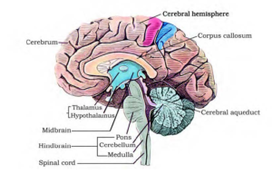
1. The brain is the central information processing organ of the body acting as the ‘command and control system’. It is protected in the skull. 2. It is covered by three membranes known as cranial meninges – the outer layer is the dura mater which is a fibrous and a tough membrane, the middle layer is the arachnoid, which is delicate and thin, the innermost layer is the pia mater which is an extension of the brain tissue. This layer is extremely vascular and supplied richly with blood 3. The three main regions of the brain are: (i) Forebrain (ii) Hindbrain (iii) Midbrain Forebrain – has three main parts – cerebrum, hypothalamus, thalamus →Cerebrum forms the most important and major part of the entire brain. It is longitudinally segregated into halves by a deep cleft, each half is known as the cerebral hemisphere. Both these hemispheres are linked by the corpus callosum which is a tract of nerve fibers. The cerebral hemispheres are internally hollow and the walls of the cerebrum have an inner medulla and the outer cortex. The cerebral cortex consists of cell bodies of neurons which imparts the grey appearance; hence it is referred to as grey matter. The grey matter has many grooves (sulci) and folds (gyri). Higher the number of convolutions, greater the intelligence. The cerebral cortex consists of sensory areas, motor areas and association areas (neither motor nor sensory). These specific areas are responsible for the complex functions namely communication, memory and intersensory associations. The cerebral medulla is made of axons of nerve fibers, imparts a white appearance, hence it is referred to as white matter. There a group of interrelated deep structures inside the cerebral hemispheres, namely the amygdala and hippocampus which results in the formation of a complicated structure known as the limbic system or the limbic lobe. Role – The cerebrum is the centre of memory, intelligence, consciousness, voluntary actions and will power →Thalamus It is made up of grey matter and located superior to the midbrain. Role – it relays motor and sensory impulses to the cerebrum and also controls the manifestation of emotions, comprehends heat, pain and cold. →Hypothalamus Located at the base of the thalamus, it consists of the optic chiasma. It is a point wherein the optic nerve fibers cross opposite sides. Behind this structure is the infundibulum, which is a greyish protuberance of the hypothalamus. It contains the pituitary gland. Role – The hypothalamus has centres responsible in regulating temperature of the body, hemeostasis, blood pressure, centre to control appetite (hunger, sleep, fatigue, thirst, pleasure, anger and penance). The neurosecretory cells of the hypothalamus produce releasing factors or several hormones that are crucial in regulating the activities of the pituitary hormones. Along with the limbic system, the hypothalamus also plays a part in regulating the sexual behavior. Midbrain It consists of the cerebral peduncles and the corpora quadrigemina →Cerebral Peduncles They are fibrous thick tracts which connect the cerebrum and the cerebellum. Role – Relay the sensory and the motor impulses between the hindbrain and the forebrain →Corpora quadrigemina The dorsal part of the brain has two pairs of solid lobes which are referred to as the corpora quadrigemina where one pair is referred to as the superior colliculi and the other pair is referred to as the inferior colliculi Role – Corpora quadrigemina controls the visual reflexes and the movement of the eye and head. They also regulate auditory reflexes and movement of the head to identify and detect the source of sound. Hindbrain It consists of the cerebellum, pons varolii and medulla oblongata →Cerebellum Present behind the top part of the brain stem. The outer cerebellar cortex consists of grey matter and the inner cerebellar medulla consists of white matter. The cerebellum is connected with the medulla oblongata and the cerebrum through the fiber tracts of the white matter. Role – It coordinates the balance of the body and the muscular activity. The impulse of the performing muscular activity is initiated in the cerebrum. It controls the voluntary movements originating in the cerebrum. →Pons varolii It is built of a thick bundle of white nerve fibers that is found above the medulla oblongata. Role – Synchronizes between both the lobes of the cerebellum. It has the center to control breathing which is referred to as the pneumotaxic center. →Medulla oblongata It is conical in shape and is located at the skull’s base. It runs behind the brain as the spinal cord. Any injury to this site of the brain could be fatal. Role – serves as a passage to conduct nerve impulses from the spinal cord to the brain. It controls all the activities of the internal organs, breathing and heartbeat. (b) Structure of the Eye –
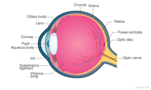
Human eye is embedded in a bony socket of the skull and they are spherical. The walls of the eye ball consist of three layers, namely – The inner neurosensory coat, the middle vascular coat and the outer fibrous coat. Outer fibrous coat – it is a thick and tough covering protecting the eye ball and helping to maintain its form. It has two regions – cornea and sclera Sclera – consists of a dense white fibrous connective tissue, where only the white eye is visible, the remaining major part is orbiting. The white of the eye is composed of collagen fibers. Role – maintaining and protecting the shape of the eyeball. Cornea – It is the non-vascular transparent part of the outer fibrous coat that is visible and is covered by a thin, transparent vascular layer of stratified epithelium known as the conjunctiva. It is in the continuation with the lining of the eyelids. Role – The cornea refracts light which enters the eye and converges it into the lens. Middle vascular coat – it consists of three regions, namely – choroid, ciliary body, and iris. Choroid – Highly vascular and made of loose fibrous connective tissue. It is in the continuation on the inner portion of the sclera. It finely layers over the posterior two-thirds of the eye ball and tends to turn thicker towards the front, imparting a bluish appearance. It consists of some pigmented cells. Role – nourishes the retina supplying it with oxygen. The pigmented cells absorb excessive light to avoid reflection in the eye ball. Ciliary body – It is thick and forms the anterior part of the choroid. Comparatively, it is less pigmented and vascular and is composed of ciliary muscles and ciliary processes. →Ciliary muscles – They are smooth muscles and are of two types – circular muscles and meridional muscles. →Ciliary processes – the inner portion of the ciliary body has plenty of folds known as ciliary processes. Role – secrete aqueous humour. Iris – it is a fine, opaque and a pigmented structure, located at the junction of sclera and cornea. The color of the iris is imparted by the pigmented cells of the choroid. The color varies between black, dark-brown, blue or green. It consists of a pupil centrally, as an aperture. The iris consists of two types of smooth muscles – circular muscles and the radial muscles. Role – iris controls the eye size and hence the amount of light that enters. When radial muscles contract, pupil enlarges in dim light. When circular muscles contract, the pupil diminishes in bright light Inner neurosensory coat – The retina forms the innermost, neurosensory fine layer of the eyeball. The outer surface of retina is in contact with the choroid and the inner surface is in contact with the vitreous humor. The external surface has four layers: Pigmented layer – it is made up of single layer of cells containing dark-brown pigment. The layer of photoreceptors – has two types of cells – rods and cones Rods – it is rod-shaped and elongated containing a purplish-red protein pigment known as rhodopsin or visual purple. It contains vitamin A derivatives. Rods do not respond to colours and are sensitive to dim light. They provide vision in the dark, hence known as twilight vision or scotopic vision Cones – these are sensitive to colors and bright lights, providing daylight or photopic vision. The pigment that is found in the cone cells is known as iodopsin. Three kinds of cone cells respond to green, red and blue light. Other colors are detected by the simultaneous trigger of cone cells of more than one kind. A sensation of white light is generated when all the three types of cells are simultaneously triggered. Cone cells are insensitive to the dim light hence in the dark, colour cannot be recognized. Layer of bipolar neurons, layer of ganglionic cells are the two layers. This layer contains the cell bodies of the ganglion cells which forms the optic nerve. Blind spot – The optic nerve exits the brain, retinal blood vessels enter the brain at a point where the photoreceptor cells are absent, this is the blind spot. Macula lutea – It is the yellowish pigmented spot that is present lateral to the blind spot. It is located exactly opposite the cornea’s centre. It has a pit located centrally known as the fovea which lacks in rods and blood vessels. It has only cone cells and is the area of most distinct vision. Lens – It is elastic, transparent and biconvex in nature which is located just behind the iris. It is covered by a thin, elastic and transparent membrane known as the lens capsule. The lens is intact in its position due to the suspensory ligaments. These ligaments along with the lens segregates the eye ball into two chambers known as the aqueous chamber and the vitreous chamber. Aqueous chamber – it is the space between the cornea and the lens containing a thin watery fluid known as aqueous humor. Vitreous chamber – it is the space between the lens and the retina which is filled with a transparent gel known as vitreous humor. Ear
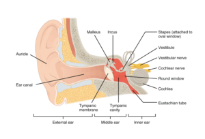

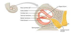
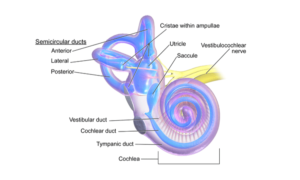
the body. It can be divided into three main sections – inner ear, outer ear, middle ear. →Inner ear – also known as the labyrinth, it is split into membranous labyrinth and the bony labyrinth. Membranous labyrinth is filled with endolymph while bony labyrinth is filled with perilymph. The Membranous labyrinth is segregated into two portions – vestibular apparatus and the cochlea. The vestibular apparatus consists of three semi-circular canals and otolith. Each semi-circular canal lies in a different plane at right angles to each other. The membranous canals are suspended in the bony canals(perilymph). The base of the canals is swollen and is known as ampulla containing crista ampullars – a projecting ridge which has hair cells. The utricle and the saccule have a projecting ridge known as macula. The macula and the crista are the particular receptors of the vestibular apparatus that has a role to play in maintaining the posture and body balance. Sacculus has a coiled and long outgrowth – cochlea which is the chief hearing structure consisting of three membranes. A hearing organ, the organ of corti, is situated on the basilar membrane possessing hair cells. →Outer ear – it has the pinna and the external auditory canal (meatus). The pinna gathers the vibrations in the air that generate sound. The external auditory canal extends up to the ear drum (tympanic membrane). It has very fine hair, wax-secreting glands in the skin of the meatus and the pinna. The tympanic membrane consists of connective tissues covered with mucous membrane inside and with skin on the outside. →Middle ear – it consists of three ossicles known as the malleus, stapes and incus that are linked to one another in a chain pattern. The malleus is linked to the tympanic membrane and the stapes is linked to the oval window of the cochlea. The ear ossicles increase the efficiency of transmission of sound waves to the inner ear. The middle ear cavity is connected to the pharynx through the Eustachian tube which aids in equalizing the pressure on both sides of the ear drum.
2. Compare the following: (a) Central neural system (CNS) and Peripheral neural system (PNS) (b) Resting potential and action potential (c) Choroid and retina
Solution: The comparison is as shown below: (a) Central neural system (CNS) and Peripheral neural system (PNS) Central neural system (CNS) Peripheral neural system (PNS) Consists of the spinal cord and the brain It consists of the spinal nerves and the cranial nerves Spinal column is protected by the vertebral column whereas the brain is protected by the skull No protective structures No subdivisions It is divided into autonomic nervous system and the somatic nervous system Processes information and regulates the responses to impulses. Nerves of PNS passes impulses to the CNS and responses from the CNS to various structures of the body Group of neurons is known as nuclei Group of neurons is known as ganglia (b) Resting potential and action potential Resting potential Action potential When the neuron is at the resting phase, it is the potential difference across membrane When the neuron is triggered it is the potential difference across the membrane The exterior side of the neuron is positively charged while the interior side is negatively charged The exterior side of the neuron is negatively charged and the interior side of the neuron is positively charged Permeability of K+ ions are observed to be more by the plasma membrane of neurons Permeability of Na+ions are observed to be more by the plasma membrane of the neurons To maintain the resting potential, the sodium-potassium ATPase pump is activated, sending Na+ions outside the neuron It functions in a reverse pattern wherein the sodium-potassium ATPase pump sends Na+ions to the neuron. (c)Choroid and retina Choroid Retina Forms the mid coat of the eyeball Forms the inner coat of the eye ball Forms the vascular layer of the eyeball Forms the neurosensory layer of the eyeball Has no photoreceptor cells Has two kinds of photoreceptors – rods and cones Prevents reflection of light in the eye and nourishes the retina Imparts vision
3. Explain the following processes: (a) Polarisation of the membrane of a nerve fibre (b) Depolarisation of the membrane of a nerve fibre (c) Conduction of a nerve impulse along a nerve fibre (d) Transmission of a nerve impulse across a chemical synapse
Solution: (a)Polarisation of the membrane of a nerve fiber:
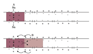
Impulse conduction through an axon A nerve fiber is said to be in a polarized state when it is at a resting phase. In this polarized state, the membrane of the nerve fibers undergo a resting potential. Listed below are the steps that occur during the process of polarization of the membrane of a nerve fiber: 1. Initially, when a depolarized region of a nerve fiber becomes polarized, the count of K+ions outside the nerve fibers is more while the axon membrane has an excessive number of Na+ions. 2. When the membrane starts to turn into a polarized state, it turns more permeable to the K+ions and impermeable to the negatively charged proteins and the Na+ions 3. The 2 K+ions are passed to the axon by a sodium-potassium pump through active transport while the 3 Na+ions are passed outside the axon 4. The outer side turns electropositive while the inner side of the membrane turns electronegative due to the movement of potassium and sodium ions which causes the nerve fiber to be polarized. (b) Depolarisation of the membrane of a nerve fibre
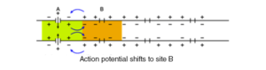
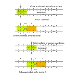
1. A nerve fiber is said to be in a depolarized state when it is triggered 2. In this state, action potential is experienced by the membrane of the nerve fiber 3. During the process of depolarization of the membrane of the nerve fiber, the following steps take place: (i) Axon has more concentration of K+ions in a polarized state and outside the axon, the Na+concentration is more. (ii) The permeability of the membranes of Na+and K+ions is reversed when the nerve fiber is triggered by the stimulus. (iii) The permeability for Na+ions by the membrane increases (iv) A rapid influx of Na+ions into the axon are observed (v) Hence, the inner side of the membrane turns positively charged while the outer side of the membrane turns negatively charged (vi) This causes depolarization of the membrane of the nerve fiber, resulting it to experience an action potential (c) Conduction of a nerve impulse along a nerve fibre 1. When a nerve impulse is conducted across the nerve fiber, it takes place in an organized manner 2. During the conduction of an impulse on the nerve fiber, a portion is depolarized always while the adjacent region is polarized. In order for the impulse to advance, repolarization of the depolarization area occurs while the polarized area depolarizes which continues across the entire length of the nerve fiber helping in the impulse conduction. 3. It takes place in the following stages: (i) Let A be a site on the depolarized region, where the inner surface of the membrane is positively charged while the outer surface of the membrane is negatively charged (ii) At site B, the adjacent region is polarized, where the outer surface is positively charged while the inner surface of the membrane is negatively charged (iii) Therefore, the flow of current at site A is on the inner surface of the membrane from A to B and at the site B, the flow of current is on the outer surface from B to A. This completes the circuit of current flow. (iv) This causes the site B to depolarize so as to conduct the impulse at site B (v) Site A gets repolarized as soon as the impulse is conducted to site B (vi) Assume a site C adjacent to site B which will be polarized when the site B is in a depolarized state.
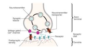
(d) Transmission of a nerve impulse across a chemical synapse

1. The membranes of the pre-synaptic neuron and the post-synaptic neuron form a synapse 2. A synaptic cleft is a synapse which may or may not be segregated by a gap 3. The pre-synaptic neuron and the post-synaptic neuron at a chemical synapse are separated by the synaptic cleft 4. The calcium ions present at the synaptic cleft enters the synaptic knobs located at the axon terminal of the pre-synaptic neuron when an impulse reaches the axon terminal 5. The synaptic knobs have the synaptic vesicles of the pre-synaptic neuron which advance towards the plasma membrane to fuse with it 6. In the synaptic cleft, the vesicles release the neurotransmitter acetylcholine 7. The acetylcholine molecules tend to bind to the protein receptors located on the plasma membrane of the post-synaptic neurons 8. The binding opens up channels for sodium ions to enter the post-synaptic neuron. Simultaneously, the potassium ions exit the post-synaptic membrane 9. This causes an action potential in the post-synaptic neuron membrane. Thus, the impulse is conveyed to the post-synaptic neuron.
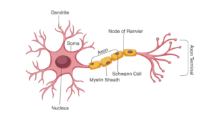
4. Draw labelled diagrams of the following: (a) Neuron (b) Brain (c) Eye (d) Ear
Solution: The diagrams are as follows: (a)Neuron
(b) Brain

(c) Eye

d)Ear

5. Write short notes on the following: (a) Neural coordination (b) Forebrain (c) Midbrain (d) Hindbrain (e) Retina (f) Ear ossicles (g) Cochlea (h) Organ of Corti (i) Synapse
Solution: (a) Neural coordination –It is a phenomena through which two or more organs interact and complement functionalities of each other through the neural system of the body. The various physiological processes that take place in the body are interlinked with each other. The neural and the endocrine system jointly are responsible to coordinate and integrate all the actions and activities of the organs such that they function in a synchronized manner. This neural system renders a systematic and organized network for a point-to-point connection for a prompt coordination. The endocrine system renders chemical integration through the hormones. (b) Forebrain – Forebrain– has three main parts – cerebrum, hypothalamus, thalamus →Cerebrum forms the most important and major part of the entire brain. It is longitudinally segregated into halves by a deep cleft, each half is known as the cerebral hemisphere. Both these hemispheres are linked by the corpus callosum which is a tract of nerve fibers. The cerebral hemispheres are internally hollow and the walls of the cerebrum have an inner medulla and the outer cortex. The cerebral cortex consists of cell bodies of neurons which imparts the grey appearance; hence it is referred to as grey matter. The grey matter has many grooves (sulci) and folds (gyri). Higher the number of convolutions, greater the intelligence. The cerebral cortex consists of sensory areas, motor areas and association areas (neither motor nor sensory). These specific areas are responsible for the complex functions namely communication, memory and intersensory associations. The cerebral medulla is made of axons of nerve fibers, imparts a white appearance, hence it is referred to as white matter. There are a group of interrelated deep structures inside the cerebral hemispheres, namely the amygdala and hippocampus which results in the formation of a complicated structure known as the limbic system or the limbic lobe. Role – The cerebrum is the centre of memory, intelligence, consciousness, voluntary actions and will power →Thalamus It is made up of grey matter and located superior to the midbrain. Role – it relays motor and sensory impulses to the cerebrum and also controls the manifestation of emotions, comprehends heat, pain and cold. →Hypothalamus Located at the base of the thalamus, it consists of the optic chiasma. It is a point wherein the optic nerve fibers cross opposite sides. Behind this structure is the infundibulum, which is a greyish protuberance of the hypothalamus. It contains the pituitary gland. Role – The hypothalamus has centres responsible in regulating temperature of the body, hemeostasis, blood pressure, centre to control appetite (hunger, sleep, fatigue, thirst, pleasure, anger and penance). The neurosecretory cells of the hypothalamus produce releasing factors or several hormones that are crucial in regulating the activities of the pituitary hormones. Along with the limbic system, the hypothalamus also plays a part in regulating the sexual behavior. (c) Midbrain It consists of the cerebral peduncles and the corpora quadrigemina →Cerebral Peduncles They are fibrous thick tracts which connect the cerebrum and the cerebellum. Role – Relay the sensory and the motor impulses between the hindbrain and the forebrain →Corpora quadrigemina The dorsal part of the brain has two pairs of solid lobes which are referred to as the corpora quadrigemina where one pair is referred to as the superior colliculi and the other pair is referred to as the inferior colliculi Role – Corpora quadrigemina controls the visual reflexes and the movement of the eye and head. They also regulate auditory reflexes and movement of the head to identify and detect the source of sound. (d) Hindbrain It consists of the cerebellum, pons varolii and medulla oblongata →Cerebellum Present behind the top part of the brain stem. The outer cerebellar cortex consists of grey matter and the inner cerebellar medulla consists of white matter. The cerebellum is connected with the medulla oblongata and the cerebrum through the fiber tracts of the white matter. Role – It coordinates the balance of the body and the muscular activity. The impulse of the performing muscular activity is initiated in the cerebrum. It controls the voluntary movements originating in the cerebrum. →Pons varolii It is built of a thick bundle of white nerve fibers that is found above the medulla oblongata. Role – Synchronizes between both the lobes of the cerebellum. It has the center to control breathing which is referred to as the pneumotaxic center. →Medulla oblongata It is conical in shape and is located at the skull’s base. It runs behind the brain as the spinal cord. Any injury to this site of the brain could be fatal. Role – serves as a passage to conduct nerve impulses from the spinal cord to the brain. It controls all the activities of the internal organs, breathing and heartbeat. (e) Retina It is the innermost layer containing layers of neural cells, namely – ganglion cells, bipolar cells and photoreceptor cells (mentioned in order from inside to the outside). The photoreceptor cells are of two types – cones and rods. Cones are responsible to impart daylight vision or colour vision whereas rods impart twilight vision. Image of an object is formed on the retina when light enters through the cornea, the lens. (f) Ear ossicles The middle ear possesses three ear ossicles known as malleus, incus and stapes that are interlinked to one another in a chain-like pattern. The malleus is in contact with the tympanic membrane, the incus with stapes and stapes inturn with the oval window of the cochlea. The ear ossicles promote and cause an increase in the efficiency of sound wave transmission to the inner ear. (g) Cochlea It is the coiled portion of the labyrinth. The membranes constituting cochlea, the basilar and reissner’s segregate the enveloping perilymph that is filled with the bony labyrinth into an upper scala vestibule and a lower scala tympani. The scala media (space within cochlea) is filled with endolymph and at the base of the cochlea, the scala vestibule terminates at the oval window whereas the scala tympani ends at the round window that opens to the middle ear.
(h) Organ of Corti Situated in the basilar membrane of the inner ear, the organ of corti is the organ of hearing. It contains hair cells that has auditory receptor cells which are inturn found in rows on the internal side of the organ. Stereo cilia are the processes that are found on the apical ends of the hair cells whereas the basal sections of the hair cells consist of synaptic contacts with afferent nerve fibers. Just above the rows of these hair cells, a smooth gelatinous layer known as the tectorial membrane is found. (i) Synapse –It is formed by the membranes of a pre-synaptic and a post-synaptic neuron, that may or may not be segregated by a gap known as the synaptic cleft. There are two types of synapses namely chemical synapses and electrical synapses.

6. Give a brief account of: (a) Mechanism of synaptic transmission (b) Mechanism of vision (c) Mechanism of hearing
Solution: (a) Mechanism of synaptic transmission: