1. Name the components of the formed elements in the blood and mention one major function of each of them.
Solution: The components of the formed elements in blood are as follows: Erythrocytes or Red blood cells – They carry oxygen and contain the pigment, haemoglobin. Haemoglobin reacts with oxygen to form Oxyhaemoglobin that carries oxygen to areas that are deprived of oxygen in the body. Leucocytes or white blood cells – Lymphocytes are known to synthesize antibodies that neutralizes or kills germs. Neutrophils acts as a defense mechanism against bacteria known as phagocytosis. Thrombocytes or blood platelets – They aid in coagulation of blood
2. What is the importance of plasma proteins?
Solution: Significance of plasma proteins: Some of the plasma proteins are Globulins – They are involved in the defense mechanism of the body, and are also referred to as immunoglobulins Albumins –They aid in maintaining the osmotic balance of the body Fibrinogens – it plays a significant role in blood coagulation
3. Match Column I with Column II : Column I Column II (a) Eosinophils (i) Coagulation (b) RBC (ii) Universal Recipient (c) AB Group (iii) Resist Infections (d) Platelets (iv) Contraction of Heart (e) Systole (v) Gas transport
Solution: Column I Column II (a) Eosinophils (iii) Resist Infections (b) RBC (v) Gas transport (c) AB Group (ii) Universal Recipient (d) Platelets (i) Coagulation (e) Systole (iv) Contraction of Heart
4. Why do we consider blood as a connective tissue?
Solution: Blood is a connective tissue as it is mesodermally derived and contains an extra-cellular matrix known as plasma. It is abundant and a widely distributed tissue in the body. Connective tissues link and bind, providing support to other organs of the body thereby transporting oxygen and other nutrients within the body, eliminating waste products from the body and flows throughout the body. Hence, it is considered as a connective tissue.
5. What is the difference between lymph and blood?
Solution: The difference between lymph and blood is as follows: Lymph Blood It is a white tissue fluid It is a red liquid connective tissue Lymph flows in the lymph vessels Blood flows in the blood vessels such as capillaries, arteries and veins It contains white blood cells known as lymphocytes Blood contains red blood cells, haemoglobin, platelets and white blood cells The exchange of nutrients and gases between the blood and cells takes place through the lymph Blood transports gases and other nutrients to the body.
6. What is meant by double circulation? What is its significance?
Solution: Double circulation, as the name suggests, is where the blood circulates twice in the heart. Double circulation is possible as the heart is divided into four chambers, the right and the left halves by the atrio-ventricular septum. The two circulations are: 1. Pulmonary circulation Blood in the right ventricle is pumped into the pulmonary arteries For oxygenation, these pulmonary arteries transport deoxygenated blood to the lungs The oxygenated blood is then sent to the left atrium from the lungs through the pulmonary veins This type of circulation of blood is referred to as pulmonary circulation where blood is pumped via pulmonary blood vessels. 2. Systemic circulation It is a term used to refer to the major circulation of the body Oxygenated blood is pumped from the left ventricle into the aorta Furthermore, it is carried by the arteries, arterioles and the linkage of blood capillaries Simultaneously, deoxygenated blood is accumulated in the right atrium through the inferior and superior vena cava This circulation supplies nutrients and oxygen and carries away carbon dioxide and other toxic substances for elimination Importance of double circulation: This type of circulation checks and prevents the mixing of oxygenated and deoxygenated blood In double circulation, oxygen is utilized efficiently
7. Write the differences between: (a) Blood and Lymph (b) Open and Closed system of circulation (c) Systole and Diastole (d) P-wave and T-wave
Solution: The differences are as follows: (a) Blood and Lymph Lymph Blood It is a white tissue fluid It is a red liquid connective tissue Lymph flows in the lymph vessels Blood flows in the blood vessels such as capillaries, arteries and veins It contains white blood cells known as lymphocytes Blood contains red blood cells, haemoglobin, platelets and white blood cells The exchange of nutrients and gases between the blood and cells takes place through the lymph Blood transports gases and other nutrients to the body. (b) Open and Closed system of circulation Open system of circulation Closed system of circulation Blood that is pumped by the heart passes through the large vessels into the open spaces or body cavities(sinuses) Blood pumped by the heart flows via the closed network of the blood vessels Blood flow is not regulated in this type of circulation Blood flow is regulated by the valves in closed system of circulation This circulation is slower and less efficient This circulation is more rapid and efficient comparitively Example – it is found in molluscs, arthropods Example – it is found in chordates and annelids (c) Systole and Diastole Systole Diastole Systole is the contraction of the chambers of the heart Diastole is the relaxation of the chambers of the heart It causes an increase in the blood pressure within the heart It causes the blood pressure to decline in the heart Blood is pumped out of the chambers Blood is received by the chambers (d) P-wave and T-wave P-wave T-wave P-wave depicts the depolarization or electrical excitation of the atria T-wave depicts the repolarization of the ventricles Blood is pumped into the ventricles Blood is received by the atria
8. Describe the evolutionary change in the pattern of heart among the vertebrates.
Solution: An evolutionary change in the pattern of heart among the vertebrates has been observed through careful analysis. Vertebrates possess a muscular heart, it is chambered. They have evolved from having a two-chambered heart(fish) to possessing a four-chambered heart(mammals). A fish has a two-chambered heart. It pumps deoxygenated blood to the gills where it is oxygenated and sent to the body. The blood that is oxygenated is then carried to the heart. Three-chambered hearts are found in amphibians – a ventricle and 2 atria(left atrium and right atrium). The left atrium receives oxygenated blood from the respiratory organs while deoxygenated blood is received by the right atrium from the organs of the body. But, both types of blood are eventually mixed in the ventricle, hence the body receives mixed blood. Half septum in reptiles divides the ventricle partially. But in birds, crocodiles and mammals, the heart is completely segregated into halves hence deoxygenated and oxygenated blood are separated. A structural modification in the hearts of fish up till mammals is observed checking that oxygen-rich blood is supplied to the body while the four-chambered heart ensures that the blood flow is synchronized. As the structure of heart has evolved, the type of circulation also depends on it, if it is single or double circulation.
9. Why do we call our heart myogenic?
Solution: The term ‘myo’ refers to muscle and ‘genic’ refers to originating from. The altered cardiac muscles or the nodal tissues of the heart – the sino-atrial or the sinus node (SA node) is capable of generating an impulse that extends over the heart wall which results in a heartbeat. As the cardiac impulse initiates in the cardiac muscles, it is referred to as myogenic.
10. Sino-atrial node is called the pacemaker of our heart. Why?
Solution: The sino-atrial or the sinus node (SAN) is a specialized bundle of neurons generating action potential which produces a cardiac impulse without any exterior stimuli i.e., it is auto excitable. SAN can generate a maximum action potential of approximately 70 to 75 in a minute. It is responsible to initiate and maintain the rhythmic contractile activity of the heart. Due to these capabilities, SAN is referred to as the pacemaker.
11. What is the significance of atrio-ventricular node and atrio-ventricular bundle in the functioning of heart?
Solution: The atrio-ventricular bundle (AV) originating from the AV-node passes the cardiac impulse to the walls of the ventricles whereas the atrio-ventricular node(AVN) conveys the impulse from the SA node. Both AVN and AV are triggered by the action potential that is originated by the sino-atrial node thereby conducting the stimulus to the other parts of the heart. This causes a heart beat which is conducted to various other parts of the heart.
12. Define a cardiac cycle and the cardiac output.
Solution: Cardiac cycle: The alternate contraction and relaxation of the chambers of the heart causes the blood to circulate in the heart. Relaxation or expansion phase is also referred to as diastole whereas contraction is also referred to as systole Every systole is followed by a diastole Cardiac cycle is the series of events that take place in one complete heart beat. It lasts for about 0.8 seconds Cardiac output: It is the amount of blood that is pumped by each of the ventricle in a minute It is given by: Cardiac output = stroke volume x number of heart beats per minute
13. Explain heart sounds.
Solution: A heart beat generates two sounds – lub and dub, produced in every cardiac cycle. The sound is produced in a sequence, in-sync with every heartbeat. ‘Lub’ is the first sound and is low pitched. It is caused when the bicuspid valve and the tricuspid valve closes. ‘Dub’ is the second sound which arises when the semi-lunar valve closes. This sound is high- pitched. Both the sounds are significant in the diagnosis of any disorder of the heart.
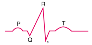
14. Draw a standard ECG and explain the different segments in it.
Solution: The graphical representation of the cardiac cycle that is generated by the electrograph is known as the electrocardiogram or ECG. The diagram below depicts a standard ECG: