Chapter 20 – Locomotion and Movement Questions and Answers: NCERT Solutions for Class 11 Biology

Class 11 Biology NCERT book solutions for Chapter 20 - Locomotion and Movement Questions and Answers.

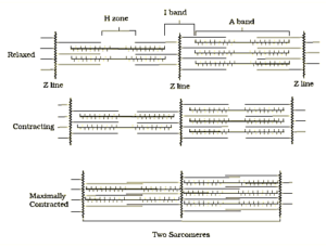
1. Draw the diagram of a sarcomere of skeletal muscle showing different regions.

Solution:
The diagram below shows the sarcomere of skeletal muscle showing different regions:

2. Define sliding filament theory of muscle contraction.

Solution:
Sliding filament theory states that the muscle fibre contraction occurs due to the sliding of the thin filaments over thick filaments.

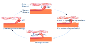
3. Describe the important steps in muscle contraction.

Solution:
The mechanism of muscle contraction is explained well by the sliding filament theory which states that the muscle fibre contraction occurs due to the sliding of the thin filaments over the thick filaments.

Mechanism of muscle contraction:
Step 1 – Depolarization of Sarcolemma
1. The central nervous system (CNS) sends a signal through a motor neuron which arrives at the neuromuscular junction or the motor end plate
2. The junction present between the sarcolemma of the muscle fiber and the motor neuron is called the neuromuscular junction
3. The release of a neurotransmitter called acetylcholine is triggered when the signal arrives at the neuromuscular junction. The action potential in the sarcolemma is set by this release.
Step 2 – Release of Calcium ions
1. The sarcolemma transmits the action potential to the sarcoplasmic reticulum in order to release the calcium ions in the sarcoplasm
Step 3 – Conformational changes occurs in the Actin filaments
1. The calcium ions that are released, tends to bind to the troponin and tropomyosin located on the active filaments
2. This linkage causes a modification in the three-dimensional structure of the actin-troponin-tropomyosin complex. The active site for myosin which is located on the actin filament is exposed.
Step 4 – Activation of Myosin heads
1. The calcium ions release causes the myosin heads to get activated which inturn causes the release of energy in the form of ATP
2. Hydrolysis of ATP releases energy which causes the binding of myosin heads to the active sites found on the actin filaments hence forming a cross-bridge
Step 5 – Actin Filaments slide over Myosin
1. The myosin head rotates due to the formation of cross-bridge, pulling the actin filaments to the centre of the A-band which is the H-zone
2. The z-line which is attached to the actin filaments is pulled inwards too
3. Contraction of sarcomere occurs when the actin filaments are pulled in the opposite ends
4. During the process of contraction, the I-band shortens whereas the A-band maintains its length causing the muscles to contract.

4. Write true or false. If false change the statement so that it is true.
(a) Actin is present in thin filament
(b) H-zone of striated muscle fibre represents both thick and thin filaments.
(c) Human skeleton has 206 bones.
(d) There are 11 pairs of ribs in man.
(e) Sternum is present on the ventral side of the body.

Solution:
(a) Actin is present in thin filament – True
(b) H-zone of striated muscle fibre represents both thick and thin filaments – False
Corrected statement: H-zone of the striated muscle fiber represents only thick filaments
(c) Human skeleton has 206 bones – True
(d) There are 11 pairs of ribs in man – False
Corrected statement: There are 12 pairs of ribs in man.
(e) Sternum is present on the ventral side of the body – True

5. Write the difference between:
(a) Actin and Myosin
(b) Red and White muscles
(c) Pectoral and Pelvic girdle

Solution:
The differences are as follows:
a) Actin and myosin
Actin
Myosin
Thin filaments (I-bands) of the myofilament are formed
Thick filaments (A-bands) of the myofilament are formed
It is built of globular actin monomers
It is built of meromyosin monomers
The regulatory proteins Tropnin and tropomyosin are linked with the actin
The regulatory proteins are not linked, instead each of the meromyosin is built of four light chains and two identical heavy chains
b) Red and white muscles
Parameters
Red muscles
White muscles
Myoglobin content
High
Low
Amount of sarcoplasmic reticulum
Moderate
High
Fibers
Narrow and thin
Broad and thick
Mitochondria
Possess many
Few
Fatigue
Not fatigued
Fatigued
Type of respiration from which energy is derived
Aerobic
Anaerobic
c) Pectoral and Pelvic girdle
Pectoral girdle
Pelvic girdle
Pectoral girdle is called as the shoulder girdle
Pelvic girdle is called as the hip girdle
Each part/half of the girdle consists of two bones – clavicle and scapula
Each part/half of the girdle consists of three bones – ischium, ilium and pubis
Forelimb articulation is offered
Hindlimb articulation is offered
Head of the humerus articulates with the glenoid cavity of the pectoral girdle
Head of the femur articulates with the acetabulum of the pelvic girdle

6. Match Column I with Column II:
Column I
Column II
(a) Smooth muscle
(i) Myoglobin
(b) Tropomyosin
(ii) Thin filament
(c) Red muscle
(iii) Sutures
(d) Skull
(iv) Involuntary

Solution:
Column I
Column II
(a) Smooth muscle
(iv) Involuntary
(b) Tropomyosin
(ii) Thin filament
(c) Red muscle
(i) Myoglobin
(d) Skull
(iii) Sutures

7. What are the different types of movements exhibited by the cells of human body?

Solution:
Movement is considered to be one of the fundamental characteristics which is observed in the living entities. The different types of movements exhibited by the human body cells are:
1. Amoeboid Movement – Leucocytes found in blood exhibit this type of movement. Leucocytes from the circulatory system move towards the injury site when there is a damage to the tissue in order to initiate an immune response.
2. Muscular movement – The muscle cells exhibit this type of movement
3. Ciliary movement – Sex cells (sperms and ova) exhibit this type of movement. This movement facilitates the passage of ova via the fallopian tube on its way to the uterus.

8. How do you distinguish between a skeletal muscle and a cardiac muscle?

Solution:
The differences between a skeletal muscle and a cardiac muscle is as follows:
Skeletal muscle
Cardiac muscle
Linked to the primary bones
Located in the walls of the heart
Voluntary in nature
Involuntary in nature
Peripherally located nucleus
Centrally located nucleus
The muscle fibers of the skeletal muscles are unbranched
The muscle fibers of the cardiac muscles are branched
Intercalated discs are not found in the Muscle fibers of the skeletal muscles
Intercalated discs are found in the muscle fibers of the cardiac muscles
Brings about the locomotory actions of the body and maintains body posture
Responsible for the movement/motion of the heart

9. Name the type of joint between the following:-
(a) atlas/axis
(b) carpal/metacarpal of thumb
(c) Between phalanges
(d) femur/acetabulum
(e) Between cranial bones
(f) Between pubic bones in the pelvic girdle

Solution:
The types of joints are as follows:
(a) atlas/axis – Pivot joint
(b) carpal/metacarpal of thumb – Saddle joint
(c) Between phalanges – Hinge joint
(d) femur/acetabulum – Ball and socket joint
(e) Between cranial bones – Fibrous joint
(f) Between pubic bones in the pelvic girdle – cartilaginous joint

10. Fill in the blank spaces:
(a) All mammals (except a few) have __________ cervical vertebra.
(b) The number of phalanges in each limb of human is __________
(c) Thin filament of myofibril contains 2 ‘F’ actins and two other proteins namely____ and _____.
(d) In a muscle fibre Ca++ is stored in __________
(e) __________ and __________ pairs of ribs are called floating ribs.
(f) The human cranium is made of __________ bones

Solution:
(a) Seven
(b) Fourteen
(c) Troponin, tropomyosin
(d) Sarcoplasmic reticulum
(e) Eleventh, twelfth
(f) Eight